 English
 Français
 Español
 Deutsch
 Italiano
 العربية



Teléfono
:+86-755-86961139
Correo electrónico
:sales@vivo-light.com

Peripheral intravenous (IV) cannulation can be challenging in patients with edema, particularly when fluid accumulation makes superficial veins difficult to identify through visual inspection and palpation. These challenges may increase the difficulty of vein selection and require additional time and attempts during venipuncture.
Previous clinical literature has explored different approaches to vascular visualization-assisted venipuncture. These include infrared vein imaging, optical vein visualization, and other visualization-assisted techniques. A review by Cai Yulan discussed the clinical applications of various visualization technologies in venipuncture, highlighting their role in supporting vein identification and puncture-site selection [5].
The 2016 Infusion Therapy Standards of Practice, published by the Infusion Nurses Society [8] and interpreted for Chinese readers by Li Chunyan [2], also addressed the use of vascular visualization technology when peripheral venous access is difficult or an initial puncture attempt is unsuccessful.
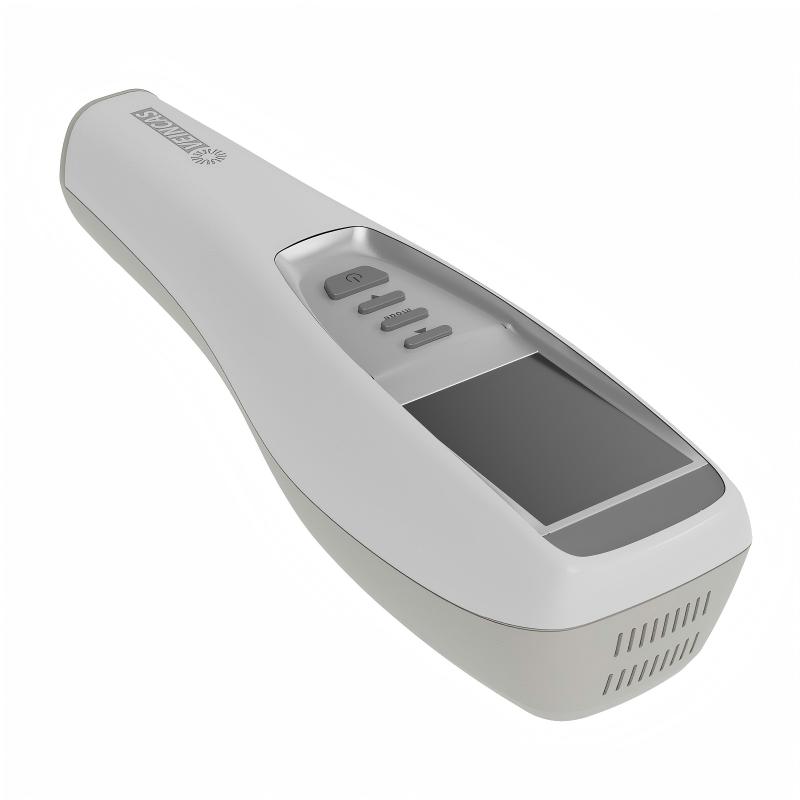
Against this background, a clinical study published in Jilin Medical Journal in December 2020 investigated the use of projection infrared vein imaging-assisted venipuncture among patients with edema. The study evaluated a projection infrared vein imaging device — the same category of device as the VIVOLIGHT projection infrared vein imaging system.
Edema occurs when excess fluid accumulates in the interstitial spaces, potentially affecting the visibility and palpability of superficial veins. In patients with significant edema, conventional vein assessment may become more challenging, making it difficult for healthcare professionals to identify an appropriate puncture site.
This issue is particularly relevant in clinical environments where nurses regularly perform peripheral IV cannulation in patients with complicated vascular conditions.
Previous studies have examined the application of projection infrared vein imaging in different patient populations. Research by Shen Ruihong et al. investigated its use in neonatal peripheral venipuncture [3], while He Ying et al. evaluated its application in pediatric peripheral venipuncture [6].
Other research has also explored high-precision infrared vein finder imaging for venipuncture in general clinical practice [4] and vascular visualization-assisted positioning techniques in elderly patient care [7].
These publications demonstrate that vascular visualization has been investigated across several clinical settings. However, the findings from different patient populations and studies should be interpreted separately rather than assumed to represent identical clinical outcomes.
The study by Chen Wanli and Li Jiexia was conducted at Guigang People's Hospital, the Eighth Affiliated Hospital of Guangxi Medical University.
Between January and December 2018, researchers enrolled 100 patients with edema who required peripheral intravenous catheterization in the nephrology department.
Patients were randomly assigned to two groups, with 50 patients in each group.
The study compared two venipuncture approaches:
Conventional venipuncture group
Nurses used traditional techniques to identify veins through clinical experience, visual inspection, and palpation before performing peripheral IV catheterization.
Visualization-assisted group
Nurses used a projection infrared vein imaging device to visualize peripheral veins, assess their course and depth, and select an appropriate puncture site.
All participating nurses were young nurses with less than five years of working experience. They received training in both venipuncture methods before the study.
The researchers evaluated two primary outcomes: first-attempt venipuncture success rate and the time required to complete the procedure [1].
The study reported two primary outcome measures — the first-attempt venipuncture success rate and the time required to complete the procedure.
First-attempt success is an important indicator when evaluating peripheral IV cannulation techniques, particularly in patients whose veins are difficult to locate.
According to the study, the first-attempt success rates were:
The difference was statistically significant (P < 0.05).
The distribution of successful attempts also showed differences:
These findings indicate that the visualization-assisted group achieved a higher first-attempt success rate under the conditions evaluated in this study [1].
The results suggest that projection infrared vein imaging may provide useful visual information to support vein identification and puncture-site selection, especially when edema makes conventional vein assessment more difficult.
The researchers also compared the time required to complete venipuncture in both groups.
The reported average procedure times were:
The difference was statistically significant (P < 0.001).
On average, the visualization-assisted group completed the procedure approximately 1.54 minutes faster than the conventional group.
This finding suggests that projection infrared vein imaging may help reduce the time spent locating suitable veins and performing peripheral IV cannulation in the patient population studied [1].
However, actual procedure times may vary depending on vascular conditions, patient characteristics, operator experience, and clinical workflow.
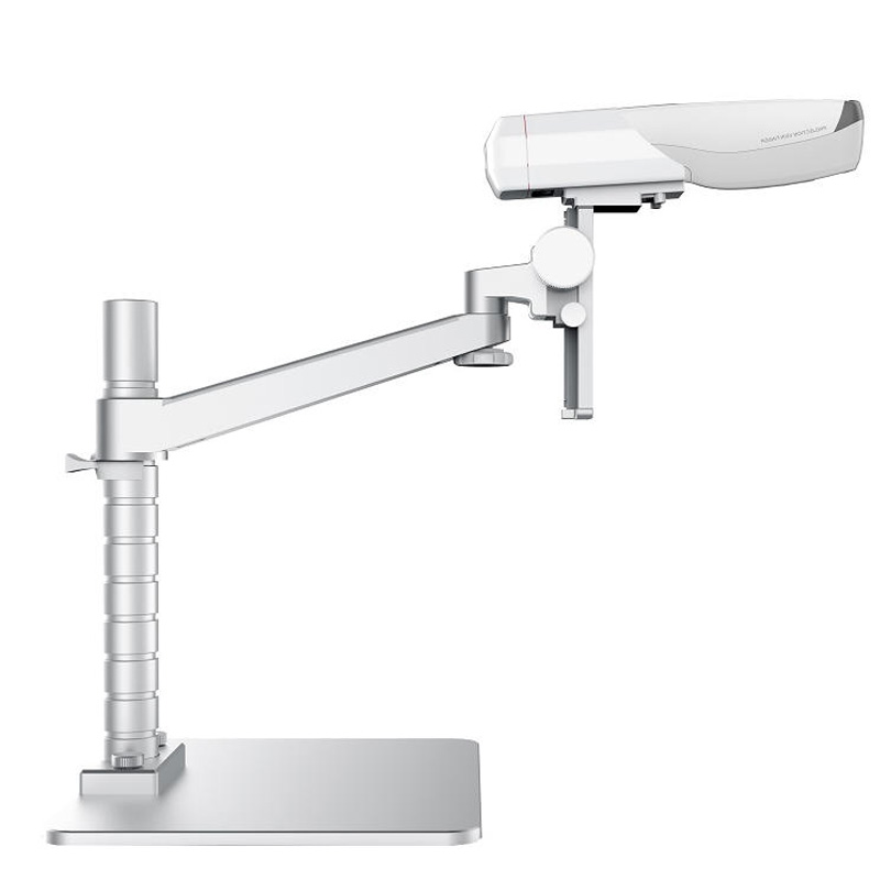
Projection infrared vein imaging is based on the different light absorption characteristics of blood and surrounding tissues.
As described in the original clinical study, hemoglobin absorbs near-infrared light differently from surrounding tissues. Digital image processing can therefore be used to generate a vascular image and project the visible vein pattern onto the skin surface.
This provides healthcare professionals with an additional visual reference when assessing superficial vein location and course.
Cai Yulan's review of clinical visualization-assisted venipuncture also discussed different approaches to vascular visualization, including vein imaging and other optical and imaging-based techniques [5].
In the study, nurses used the projected vascular image to assess vein location, size, and direction. The device's depth mode was also used to assist with evaluating vessel depth before selecting a puncture site.
The original paper reported that the device was positioned approximately 20–30 cm above the patient's skin during the procedure. Nurses could adjust the imaging mode to obtain a clearer vascular image when necessary [1].
Importantly, vascular visualization provides supplementary information rather than replacing clinical assessment, palpation, or professional judgment.
The clinical literature cited in the original study has explored vascular visualization in several patient populations.
Neonatal peripheral venipuncture
Shen Ruihong et al. investigated the application of projection infrared vein imaging in neonatal peripheral venipuncture [3]. This provides an example of research exploring visualization-assisted techniques in a patient population where vascular access can present particular challenges.
Pediatric peripheral venipuncture
He Ying et al. examined the application of projection infrared vein imaging in pediatric peripheral venipuncture [6]. Their research adds to the clinical literature on visualization-assisted vascular assessment in children.
Elderly patient care
Kou Jingli and Zhang Tong discussed vascular visualization-assisted positioning and illumination-assisted puncture techniques in elderly patient care [7]. This highlights another clinical setting in which additional visual information may be relevant to venipuncture assessment.
These studies provide broader clinical context for the development and application of vascular visualization technology. Nevertheless, their findings should not be interpreted as direct clinical validation of the VIVOLIGHT projection vein finder device unless the specific device used in each study is independently confirmed.
The 2020 study provides evidence supporting the potential value of projection infrared vein imaging-assisted venipuncture in patients with edema.
Three observations are particularly relevant:
1. Support for first-attempt venipuncture
The visualization-assisted group achieved a first-attempt success rate of 92%, compared with 72% in the conventional group. This suggests that additional vascular visualization may support more informed puncture-site selection in the specific clinical setting evaluated.
2. Improved procedural efficiency
The average venipuncture time was approximately 1.54 minutes shorter in the visualization-assisted group. This may help reduce the time required for vascular assessment and peripheral IV cannulation.
3. Additional support for less experienced nurses
Because the study involved nurses with less than five years of working experience, its findings also highlight the potential value of visualization technology as an additional reference during venipuncture training and clinical practice.
These findings should be interpreted within the study's limitations. The research involved 100 patients at a single hospital and focused specifically on patients with edema in a nephrology department. Further research involving larger populations and different clinical settings would help establish the broader applicability of these findings.
Projection infrared vein imaging offers an additional visualization approach for peripheral venous access, particularly when edema makes superficial veins difficult to identify through conventional assessment.
In a 2020 clinical study involving 100 patients with edema, a projection infrared vein imaging device was used to assist peripheral IV cannulation. The visualization-assisted group achieved a first-attempt success rate of 92%, compared with 72% in the conventional group. The average procedure time was also shorter, at 2.81 minutes versus 4.35 minutes.
Together with previous research exploring vascular visualization in neonatal, pediatric, and elderly patient care, these findings contribute to the clinical literature on visualization-assisted venipuncture.
As an adjunctive visualization tool, projection infrared vein imaging may provide healthcare professionals with additional information during vascular assessment while complementing their clinical skills and judgment.
[1] Chen W, Li J. Observation on the effect of a projection far-infrared vein imaging-guided venipuncture method in improving the venipuncture success rate of young nurses (within five years of practice) treating patients with edema. Jilin Medical Journal. 2020;41(12):3042–3044.
[2] Li CY. Interpretation of the key points of the American INS 2016 Infusion Therapy Standards of Practice. Chinese Nursing Management. 2017;17(2):150–153.
[3] Shen RH, Luo LF, Chen ZX. Application of a projection infrared vein imaging device in neonatal peripheral venipuncture. Journal of Qilu Nursing. 2018;24(4):80–81.
[4] Dai JL. Observation on the clinical effect of venipuncture using a projection infrared vein imaging device. Today Nurse. 2016;(7):141–142.
[5] Cai YL. Progress in the clinical application of visualization-assisted venipuncture. Journal of Minimally Invasive Medicine. 2015;10(6):785–788.
[6] He Y, Xiao LP, Li RJ. Application effect of a projection infrared vein imaging device in pediatric peripheral venipuncture. China Modern Medicine. 2015;22(31):192–194.
[7] Kou JL, Zhang T. Application of vascular visualization-assisted positioning and illumination-assisted puncture in elderly patient care. Nursing Research of China. 2018;32(12):1909–1911.
[8] Infusion Nurses Society. Infusion Therapy Standards of Practice. Journal of Infusion Nursing. 2016;39(1 Suppl):S1–S159.
Dejar un mensaje
Escanear a WhatsApp :


 IPv6 RED SOPORTADA