 English
 Français
 Español
 Deutsch
 Italiano
 العربية


Teléfono
:+86-755-86961139
Correo electrónico
:sales@vivo-light.com

Primer caso en los Emiratos Árabes Unidos con el sistema OCT multimodal Vivolight P80.
Dubái, Emiratos Árabes Unidos — 2 de junio de 2026
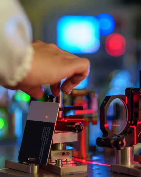
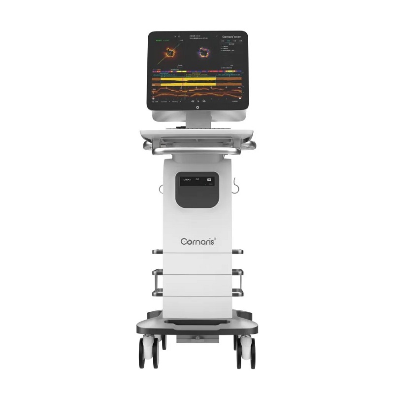
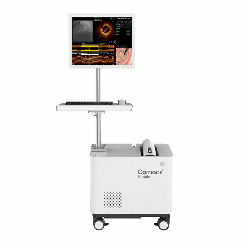
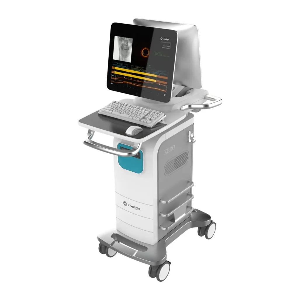
Vivolight Multimodal P80 El sistema OCT ha completado con éxito su primer caso clínico en el Hospital Aster Mankhool en los Emiratos Árabes Unidos, lo que supone un hito importante para la obtención de imágenes intravasculares avanzadas en la guía de la ICP.
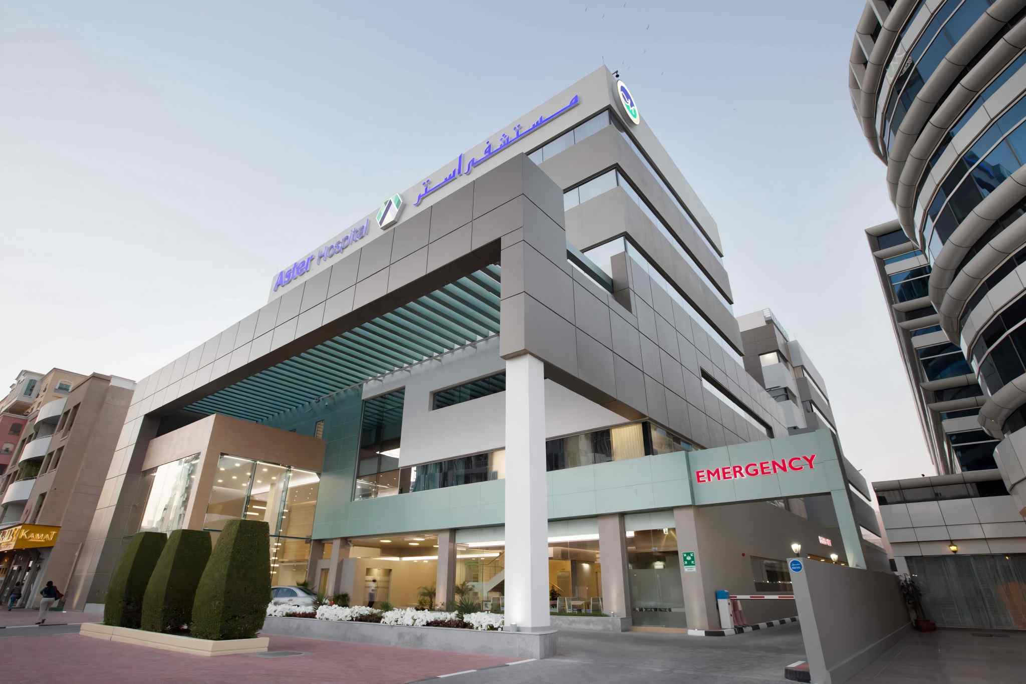
Ubicado en Dubái, el Hospital Aster Mankhool es un hospital multidisciplinario líder, reconocido por sus servicios clínicos integrales y su atención centrada en el paciente. El hospital ocupó el cuarto lugar en los Emiratos Árabes Unidos en la clasificación de los Mejores Hospitales del Mundo 2026 de Newsweek, lo que refleja su compromiso constante con la excelencia clínica y la prestación de servicios de salud de alta calidad.
La finalización exitosa de este primer caso demuestra la creciente adopción de la ICP guiada por OCT en Oriente Medio y destaca el valor de las imágenes intravasculares de alta resolución para respaldar una intervención coronaria más precisa.
Al ser el primer sistema Vivolight Multimodal P80 OCT instalado en los Emiratos Árabes Unidos, este caso representa un importante avance en la incorporación de la imagen intravascular avanzada basada en IA a la práctica rutinaria de la cardiología intervencionista.
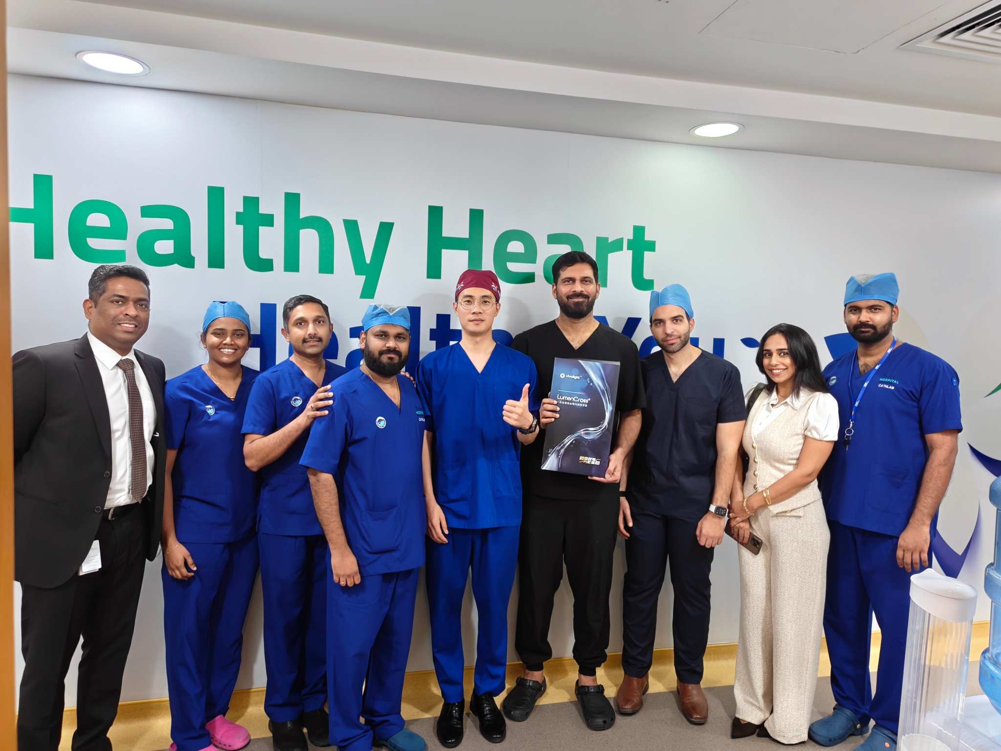
El procedimiento fue realizado por Dr. Nawid y se trató de un caso complejo de enfermedad de las arterias coronarias de tres vasos. En una de las lesiones más distales, el área luminal se midió en solo aproximadamente 1,0 mm², lo que supone un desafío significativo para la administración de catéteres de imágenes intravasculares. Gracias a la excelente capacidad de cruce Gracias al catéter Vivolight OCT, el sistema logró atravesar con éxito el segmento distal estrechado y permitió una evaluación precisa de la lesión.
Las imágenes de OCT mostraron características compatibles con una placa fibrosa, lo que proporcionó información importante para la selección de la estrategia de tratamiento, incluido el uso de un balón recubierto de fármaco. Al ofrecer una evaluación pre-PCI optimizada basada en IA de la lesión, el sistema Vivolight P80 ayudó al equipo clínico a tomar decisiones de procedimiento más informadas.
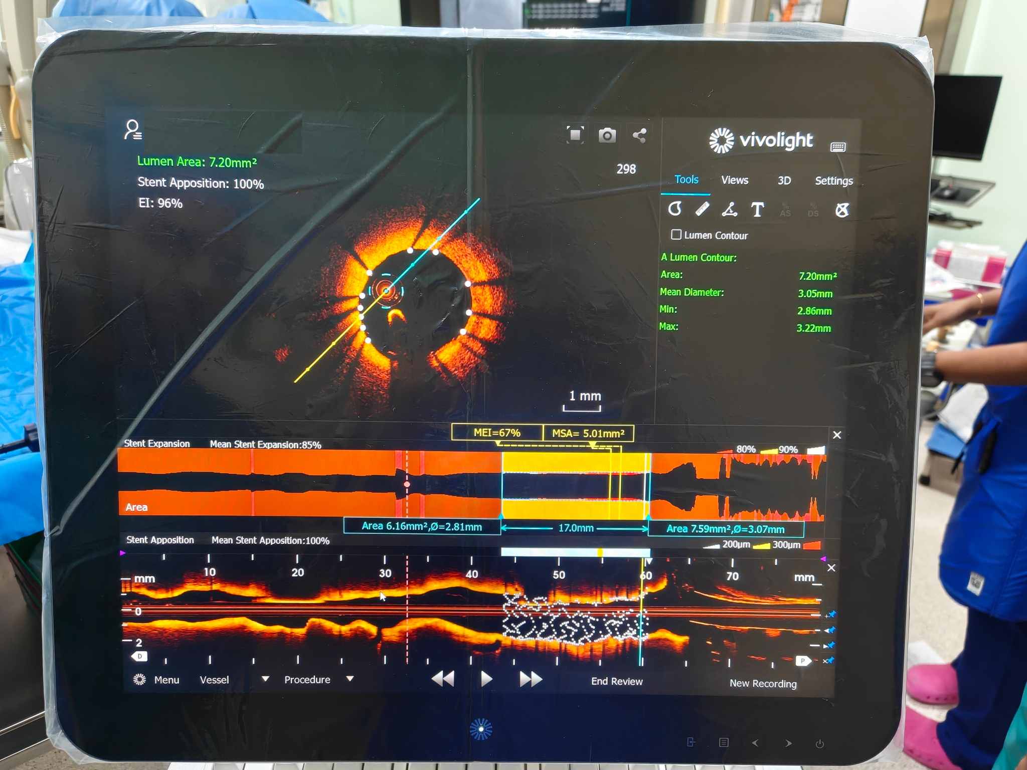
Tras la implantación del stent, se utilizó el sistema Vivolight P80 para la evaluación mediante OCT posterior a la ICP. La plataforma proporcionó vistas transversales y longitudinales de alta resolución del segmento vascular tratado, junto con un análisis cuantitativo automatizado de las dimensiones de la luz, la aposición del stent y su expansión.
En la revisión OCT posterior al procedimiento, el segmento revisado por el sistema mostró Aposición del stent al 100%, mientras que el análisis de expansión del stent mostró una expansión media del stent de 85% y un área mínima de stent de 5,01 mm²Estos datos cuantitativos obtenidos mediante OCT ayudaron al equipo clínico a evaluar el resultado inmediato del stent y demostraron el valor práctico de la ICP guiada por imágenes para la optimización del procedimiento.
El primer uso clínico exitoso del primer sistema Vivolight P80 OCT de los EAU en el Hospital Aster de Mankhool sienta una base sólida para una mayor adopción de la ICP guiada por OCT en el país.
Más allá de la obtención de imágenes intravasculares de alta resolución, el Vivolight P80 está diseñado como una plataforma OCT multimodal que integra tres tecnologías clave en un único flujo de trabajo: FFR derivada de OCT para la evaluación funcional de la estenosis coronaria, Índice de Atenuación de Placa (IPA) para la evaluación de la estabilidad de la placa y Evaluación Inteligente del Calcio (ICA) para la evaluación automatizada del calcio. Estos datos multimodales se pueden obtener a partir de una sola adquisición de OCT, lo que ayuda a los médicos a evaluar la importancia fisiológica, la morfología vascular, las características de la placa y la gravedad del calcio dentro de la misma plataforma de imágenes.
Para los procedimientos de ICP, este flujo de trabajo integrado puede facilitar la evaluación de la lesión, la selección de la estrategia de tratamiento, la preparación de la lesión, la determinación del tamaño del stent y la optimización del tratamiento. En este primer caso de UAE, el sistema demostró no solo un rendimiento de imagen nítido y una colocación fiable del catéter en una lesión distal compleja, sino también un valor práctico al facilitar la toma de decisiones precisas guiadas por imágenes.
La exitosa finalización de este caso también representa un paso importante en la expansión de Vivolight hacia los mercados internacionales de imágenes cardiovasculares de alta gama. Vivolight continuará colaborando con socios clínicos en todo Oriente Medio para ampliar el acceso a soluciones avanzadas de imágenes intravasculares y brindar una atención cardiovascular más precisa, eficiente y centrada en el paciente.
Dejar un mensaje
Escanear a WhatsApp :


 IPv6 RED SOPORTADA